
Article: Understanding Dermatofibromas and What Under-the-Scope Examination Reveals
Understanding Dermatofibromas and What Under-the-Scope Examination Reveals
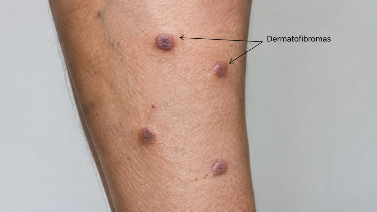
Dermatofibromas are among the most common benign skin growths that bring patients into a GP's surgery or a dermatology clinic. They are firm, often slightly raised nodules that most frequently appear on the legs, and for the majority of people who have them, they are entirely harmless. Yet their importance in clinical practice lies less in the lesion itself than in the diagnostic uncertainty it can create: because a dermatofibroma can mimic melanoma, a dermatofibrosarcoma protuberans and several other significant tumours, doctors tend to err on the side of caution when the diagnosis is unclear. Understanding what a dermatofibroma actually is — and what a dermatoscope, or the pathologist's microscope, reveals about it — makes it easier to appreciate why this quiet little lump attracts quite so much attention.
What a dermatofibroma actually is
A dermatofibroma, also called a fibrous histiocytoma, is a benign proliferation of fibrous tissue in the dermis, the deeper layer of the skin beneath the epidermis. It is thought to arise most often after minor trauma — an insect bite, a shaving nick, a thorn or an ingrown hair — which triggers a localised, over-enthusiastic healing response. Instead of resolving quietly, the area of fibrosis persists and organises into a nodule.
The typical dermatofibroma is between five and ten millimetres across, smooth or slightly scaly on the surface, and fixed to the deeper tissue so that it does not slide freely under the skin. Colour is variable: pink, tan, reddish-brown or a deeper chocolate brown. Many are darker at the periphery and paler in the centre, and some have a faintly dull, matte finish. The single most useful bedside test is the dimple sign: when the lesion is squeezed gently between finger and thumb, it sinks inward rather than popping outward, because the fibrous tissue tethers the surface to the deeper dermis. Lesions are usually solitary but may be multiple, and they occur at any age, with a peak in early to middle adulthood. They are commoner in women, largely because they favour the lower legs.
Why the diagnosis matters
The clinical problem is that dermatofibromas are chameleons. A firm, dark nodule on the leg of a fair-skinned adult could be an entirely benign dermatofibroma, or it could be a nodular melanoma, a dermatofibrosarcoma protuberans, a spitz naevus, a blue naevus, a keloid or a cutaneous metastasis. Dermatofibrosarcoma protuberans is the most important mimic, because it is a locally aggressive tumour that requires wide excision yet can look deceptively benign in its early stages. Nodular melanoma is the other great worry, since it is the subtype most likely to be thick and therefore dangerous at diagnosis.
Because the stakes of a missed malignancy are so high, no clinician should dismiss a firm nodule on sight alone. The lesion's history helps — a dermatofibroma has usually been present and stable for months or years, whereas a melanoma tends to grow and change — but history is not proof. That is where dermoscopy and, when uncertainty persists, biopsy with histopathology earn their place.
What dermoscopy reveals
Dermoscopy, or dermatoscopy, is the examination of skin lesions with a hand-held magnifier and a light source, usually with a drop of liquid or a cross-polarised filter to remove surface reflection. It allows the clinician to see structures in the epidermis and upper dermis invisible to the naked eye. For dermatofibroma, a characteristic dermoscopic picture has been well described over the past two decades.
The central feature is a central white patch — a scar-like, whitish area in the middle of the lesion corresponding to the dermal fibrosis and thickened collagen. Around this white centre, the pigment network is faint, broken and often peripheral, fading into a delicate brown ring at the edge rather than running uniformly across the whole lesion. Some lesions show fine, arborising or linear vessels at the periphery, and a few show a faint white network at the margin. When all of these elements are present together, they form a "signature" that supports the diagnosis with reasonable confidence.
The practical value of dermoscopy is that it changes management. If the pattern is classic for dermatofibroma, the clinician can often reassure the patient and avoid unnecessary surgery. If the pattern is atypical — a broad, irregular pigment network, thick irregular streaks, blue-grey veil, or asymmetric pigmentation — the lesion should be biopsied rather than watched. Dermoscopy therefore acts as a triage tool: it separates the reassuring majority from the minority who need a definitive answer.
What the microscope reveals
When a biopsy is taken, histopathology provides the definitive answer. Under the microscope, a mature dermatofibroma shows a dermal proliferation of spindle-shaped cells arranged in short, interlacing fascicles. These cells are fibroblasts and myofibroblasts — the connective-tissue cells of the dermis. Surrounding them is a variable amount of dense collagen, often in thick bundles with a characteristic whorled or storiform (cartwheel-like) arrangement. The overlying epidermis frequently shows acanthosis — a thickening of the epidermal layer — and often a central indentation or "dimpling" at the surface, which corresponds precisely to the dimple sign felt at the bedside. The lesion is well-circumscribed but not encapsulated, and it blends into the surrounding dermis rather than sitting apart from it.
Several histological variants are recognised. The cellular variant has a higher density of cells; the aneurysmal variant contains blood-filled spaces and may mimic a vascular tumour; the atrophic variant is depressed rather than raised; and the haemosiderotic variant contains haemosiderin pigment from old haemorrhage and can look dangerously like melanoma. Immunohistochemistry helps experienced pathologists distinguish these variants from their mimics — positive staining for factor XIIIa and CD34 in different patterns is used, though no single stain is perfect. Marginal excision is normally curative, and recurrence is uncommon when the lesion is completely removed.
Living with a dermatofibroma
For most patients, no treatment is needed. A dermatofibroma that has been confidently diagnosed can simply be left alone, and it often becomes less noticeable over time as the colour fades. If the lesion is symptomatic, persistently irritated by clothing, repeatedly inflamed, or cosmetically bothersome, surgical excision or shave removal can be offered — but patients should be warned that the resulting scar may look worse than the original lesion, and that recurrence is possible if the lesion is incompletely removed. Cryotherapy and laser treatment sometimes flatten the lesion but rarely eliminate it.
The most important message for anyone with a suspicious lump is simple: do not ignore change. A nodule that grows, bleeds, ulcerates, changes colour or becomes painful should be examined by a doctor rather than self-managed. Self-diagnosis by comparing images online is unreliable, and delays in presentation of a melanoma cost lives. Even where the diagnosis turns out to be a benign dermatofibroma — and happily, it usually does — the small cost of a check-up is trivial next to the reassurance it brings.
Dermatofibromas sit at an interesting crossroads in clinical dermatology. They are common, benign and generally uneventful, yet they teach a critical lesson about the limits of visual diagnosis. Dermoscopy and histology have given clinicians powerful tools to distinguish them from their far more dangerous mimics, and together they represent a tidy illustration of how careful examination — first with the eye through the dermatoscope, then with the microscope — turns uncertainty into an answer the patient and doctor can both trust.


Leave a comment
This site is protected by hCaptcha and the hCaptcha Privacy Policy and Terms of Service apply.