
Article: Spotting a Red Hand Nodule: When Is It Time to See a Dermatologist?
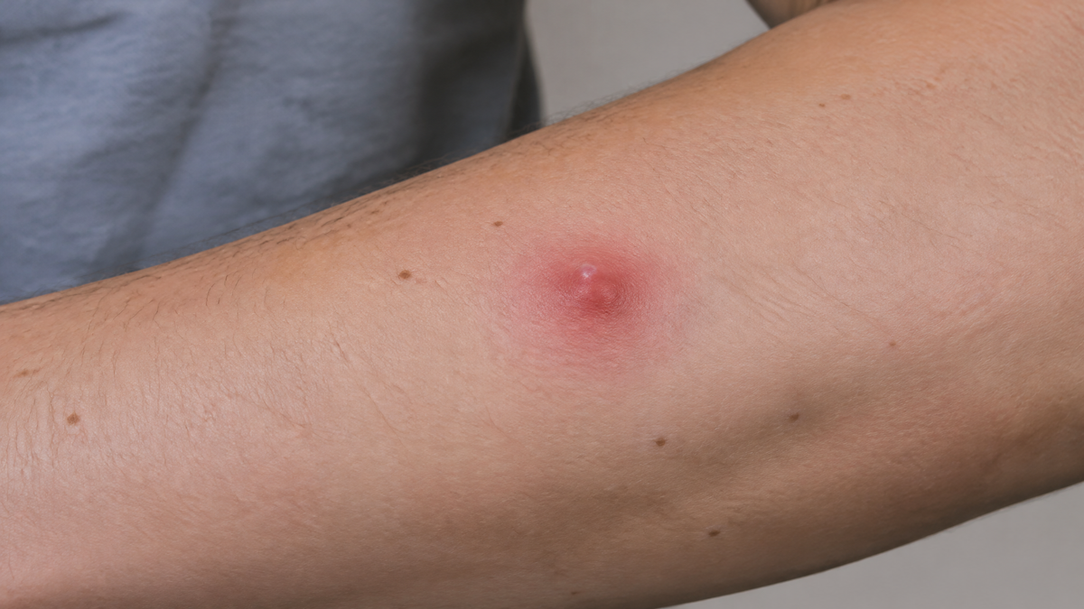
Spotting a Red Hand Nodule: When Is It Time to See a Dermatologist?
A small red lump appears on your hand. Perhaps it arrived after a knock against a doorframe, or perhaps it simply materialised on the back of a knuckle with no explanation at all. For most people the first instinct is to wait and see. That instinct is not unreasonable — the overwhelming majority of red lumps on the hands turn out to be harmless — but it is also the reason some serious skin conditions are diagnosed later than they needed to be.
The hand is a uniquely awkward place to develop a lesion. It is constantly moving, constantly exposed, constantly knocking against things, and constantly visible to you, which means you notice everything that changes and worry about all of it. This article walks through what a red nodule actually is, the conditions that commonly cause one on the hand, the specific features that should push you towards booking an appointment rather than a wait-and-see approach, and what a dermatologist will actually do when you get there.
What "Nodule" Actually Means
A nodule is a solid, raised lesion that is larger and deeper than a papule. In practical terms, dermatologists generally treat anything over roughly 5–10mm as a nodule rather than a papule, and the key distinguishing feature is depth: a nodule has substance beneath the surface rather than sitting flat against it. You can often feel a nodule as much as see it, and it may move with the skin or feel fixed to the tissue beneath.
Colour is a separate matter entirely. "Red" is a broad descriptor that spans a huge range — bright cherry red, dusky purple, violaceous, pinkish-brown, or a red so dark it looks almost black. The vascular ones tend to be red because blood is close to the surface. When you press on a red nodule and it blanches (goes pale) and then refills, that generally points towards a vascular cause. When it does not blanch, other explanations become more likely.
The Common Causes of a Red Nodule on the Hand
Pyogenic granuloma
This is one of the most frequent culprits, and it is the one that most reliably frightens people. A pyogenic granuloma — more accurately called a lobular capillary haemangioma — is a benign overgrowth of tiny blood vessels that typically appears as a bright red, fleshy, glossy bump. It grows quickly, often over a few weeks, and then stabilises. It rarely exceeds a centimetre. Its defining behaviour is spectacular bleeding from minor trauma: catching it on a sleeve or knocking it against a surface can produce a startling amount of blood for something so small.
Pyogenic granulomas appear most often on the fingers, the face and the scalp, and they are especially common in children, young adults, and during pregnancy. Minor skin injury sometimes precedes them. Importantly, a pyogenic granuloma cannot become a skin cancer — but it can closely resemble one, which is precisely why dermatologists are cautious about them. Atypical amelanotic melanoma and squamous cell carcinoma can both look like a red fleshy nodule on a hand, so a lesion assumed to be a pyogenic granuloma should still be examined by someone who knows what else it might be.
Dermatofibroma
A dermatofibroma is a firm, benign fibrous nodule that often has a reddish-brown or pink-purple tint. They frequently follow insect bites or minor injuries, and they most often appear on the lower legs, though the hands and arms are not unusual sites. The classic bedside test is the "dimple sign": pinch the nodule between thumb and forefinger and it dips inward rather than bulging outward. Dermatofibromas are harmless, but they can be itchy or tender and are occasionally removed for comfort.
Cherry angiomas
Cherry angiomas are extremely common benign vascular lesions that appear in middle age, typically as bright red or purple papules and small nodules. On a hand they read as an isolated red spot that has been there for years, unchanged. Under dermoscopy they show characteristic red or purple lacunes. They are not dangerous and need no treatment unless they bleed repeatedly or catch on things.
Chilblains (perniosis)
In cold, damp conditions — a familiar scenario in a British winter — small red or purple itchy swellings can develop on the fingers and toes. Chilblains are an abnormal vascular response to cold, and they typically resolve as the weather warms. They can, however, persist or recur, and recurrent chilblains in an unusual pattern should be assessed, as they occasionally signal an underlying condition such as lupus.
Gouty tophi and rheumatoid nodules
Not all red hand nodules are primarily skin problems. Gout can deposit urate crystals in and around the joints and tendons of the hands, producing firm nodules that may be tender or inflamed during a flare. Rheumatoid arthritis can produce rheumatoid nodules — firm, flesh-coloured or slightly reddish lumps over pressure points and joints. Both of these are managed by rheumatology, but they frequently present first as a lump the patient notices on their hand.
Infections and inflammatory causes
A red, tender, warm nodule may be infective. Abscesses, infected cysts, and deep skin infections all present this way, and they usually come with pain, warmth, and sometimes systemic symptoms such as fever. Inflammatory conditions such as granuloma annulare can also produce reddish nodules, typically in a ring pattern, though plain annular plaques are more common than true nodules.
Vascular and malignant causes
This is where the stakes rise. Squamous cell carcinoma on the hand often presents as an enlarging keratotic nodule with a raised, everted edge — a crater-like appearance. The dorsum of the hand, an area of high cumulative sun exposure, is a classic site. Keratoacanthoma looks similar and grows faster, and is considered by many dermatologists to sit on a spectrum with squamous cell carcinoma. Amelanotic melanoma — melanoma without pigment — is the lesion that hides in plain sight, and it can look like a red or pink nodule, a pyogenic granuloma, or an innocent inflamed blemish. Kaposi sarcoma and angiosarcoma are rarer, but they also enter the differential, particularly in immunosuppressed individuals.
The Red Flags: When to Book an Appointment
The following features should trigger a dermatology or GP appointment. Any single one of them is sufficient reason to be seen.
Rapid growth. A lesion that has visibly enlarged over weeks rather than months. Rapid growth is not automatically sinister — pyogenic granulomas grow fast — but it always warrants assessment.
Bleeding without clear cause. A nodule that bleeds spontaneously or from minimal trauma, or that bleeds repeatedly.
Ulceration. Any break in the surface that does not heal, or heals and then re-opens. An ulcer that has not healed within four weeks is a specific trigger for urgent referral in the UK.
Change in an existing lesion. A lump you have had for years that starts to grow, change colour, change shape, itch, or bleed.
Asymmetry and irregular borders. A lesion that is lopsided, or whose edge is ragged or poorly defined.
Colour variation within the lesion. Multiple shades — red, brown, black, blue, grey, white — within a single nodule.
Diameter. Anything over roughly 6mm is worth a professional opinion, though many benign lesions exceed this and many significant ones are smaller.
Pain or tenderness. Particularly if new, and not explained by obvious injury.
Fixation. A nodule that feels stuck to deeper tissue and does not move freely with the skin.
Satellite lesions. Small new nodules appearing around the original one.
Systemic symptoms. Fever, weight loss, night sweats, or unexplained fatigue alongside a new skin lesion.
The NHS "ABCDE" framework — Asymmetry, Border irregularity, Colour variation, Diameter over 6mm, Evolving — is designed for pigmented lesions but applies usefully to nodules too. The crucial letter for nodules is E for evolving. A lesion that is changing is the lesion that needs looking at.
If you are immunosuppressed, have a history of skin cancer, have had significant occupational sun exposure, or have a strong family history of melanoma, your threshold for seeking review should be lower than average. There is no penalty for being seen and told everything is fine.
What Happens at a Dermatology Appointment
The consultation is usually quicker and less dramatic than people expect. A dermatologist will take a history — how long the lesion has been there, how it has changed, whether it bleeds or itches, what you do for a living, your sun exposure history, your family history. Then they will look. Dermoscopy, using a magnifying instrument with polarised light, reveals structures invisible to the naked eye and can often distinguish benign vascular lesions from concerning ones on the spot.
If the diagnosis remains uncertain, or if there is any suspicion of malignancy, the next step is a biopsy. Depending on the lesion this may be a punch biopsy, an excision, or a shave. Every specimen is sent for histopathology, and histology is the only way to be certain. For a pyogenic granuloma, treatment is usually shave excision or curettage followed by cautery to the base, precisely because the diagnosis cannot be fully secured without removing it.
The reassuring truth is that most red hand nodules turn out to be benign. The reason dermatologists do not simply tell everyone to relax is that the cost of the occasional unnecessary appointment is trivial compared with the cost of a melanoma found a year late.
Where Skincare Fits — and Where It Does Not
It is worth being blunt: skincare cannot diagnose a nodule, cannot treat a malignant one, and cannot substitute for a medical opinion. No cleanser or serum will tell you whether a growing red lump is a pyogenic granuloma or a squamous cell carcinoma.
What skincare can do is look after the surrounding skin — and that matters more than it sounds. The hands take a beating. Frequent hand washing, cold weather, cleaning products and the sheer mechanical wear of daily life strip the skin barrier on the hands faster than almost anywhere else on the body, leaving it rough, cracked and slow to heal. A compromised barrier also makes the skin less resilient against environmental stressors, including ultraviolet exposure.
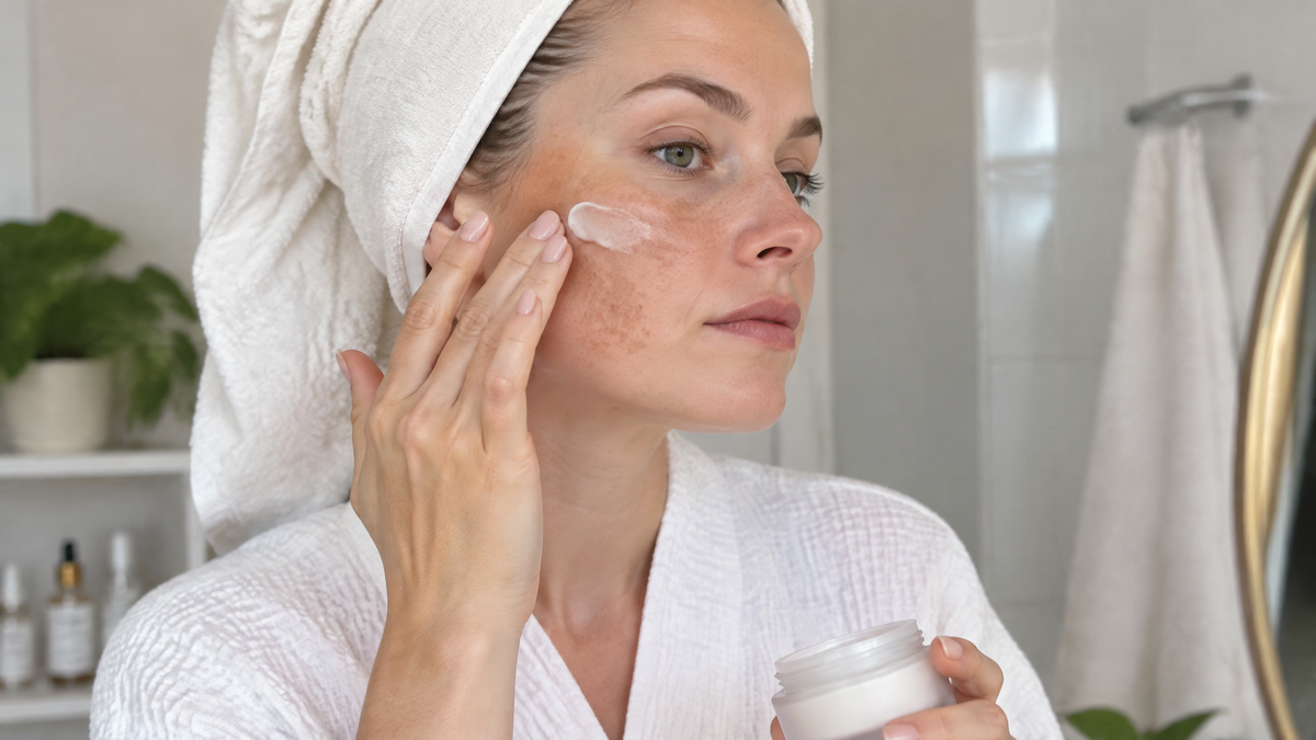
If you are managing dry, flaky or barrier-damaged hands while waiting for an appointment, gentle, fragrance-free care is the sensible route. REFORM Skincare makes a Skin Barrier Repair Cream specifically formulated as a non-greasy emollient for very dry and damaged skin, designed to support the skin's ability to bind and retain moisture and to encourage the exfoliation of rough, flaky patches — useful for hands that have been through a hard winter or a lot of washing. Their Vitamin B5 Gel is a light, fast-absorbing hydrating formula that replenishes moisture and supports the natural barrier, and is more comfortable than a heavy cream if you are applying it during the day.

Sun protection deserves a specific mention, because the dorsum of the hand is a high-exposure site and cumulative ultraviolet damage is a genuine driver of squamous cell carcinoma and melanoma there. Hands are also the area most people forget. A high-factor sunscreen applied to the backs of the hands as part of a daily routine is one of the few genuinely rational things you can do to reduce your long-term risk. REFORM's SPF 50+ Antioxidant Sunscreen and their SPF 30 Mineral Sunscreen are both options in that category.
Two important caveats. First, do not apply anything — including gentle skincare — to an open, bleeding or ulcerating lesion without medical advice; you will obscure the appearance of the lesion and potentially irritate it. Second, do not attempt to treat a nodule yourself. Over-the-counter creams marketed for lumps, home freezing kits, and "drawing salves" are all ways to delay a proper diagnosis. If you have a nodule, the correct first product is not a cream. It is an appointment.
Practical Steps While You Wait
Photograph the lesion now, with good light and something for scale such as a ruler or a coin beside it, and date the image. Take another photograph a fortnight later. This simple record gives a dermatologist far better information than your memory of "it seems a bit bigger", and it is genuinely useful evidence in a consultation.
Do not pick at it, squeeze it, or scrub it. Do not apply acids, retinoids or exfoliants to it — that is a route to inflammation, not diagnosis. Keep the area clean and covered if it bleeds, applying firm uninterrupted pressure for ten to twenty minutes if a pyogenic granuloma type lesion bleeds heavily. Protect the rest of the hand from sun exposure while you wait, and note down anything else that has changed — new symptoms, a lesion elsewhere, weight loss, night sweats.
If the nodule bleeds heavily and will not stop, or the hand becomes red, hot, swollen and painful with fever, that is an urgent situation rather than a routine appointment. Seek same-day medical care.
The Bottom Line
Most red nodules on the hand are benign, and the most common ones — pyogenic granulomas, cherry angiomas, dermatofibromas — are entirely harmless. But the hand is also a site where squamous cell carcinoma, amelanotic melanoma and other significant lesions can and do appear, and several of them mimic benign lumps closely. The features that should send you to a doctor are change: growth, bleeding, ulceration, colour variation, fixation, and a lesion that simply will not settle. A dermatologist can distinguish these in minutes with a dermatoscope, and can confirm with a biopsy when needed.
Skincare has a real role in keeping the skin around a lesion — and the hand as a whole — healthy and better protected, and REFORM Skincare's barrier and sun-protection ranges are sensible choices for that job. It has no role at all in deciding what the nodule is. That is a question for a clinician, and asking it early is the single best thing you can do.

Leave a comment
This site is protected by hCaptcha and the hCaptcha Privacy Policy and Terms of Service apply.