Full-Body Skin Cancer Screenings & Mole Mapping: A Patient Guide
Regular skin checks are one of the simplest, most powerful ways to catch skin cancer early. Yet many people put them off because they are unsure what to expect, feel embarrassed, or don’t realize they’re at risk.
This guide walks you through full-body skin cancer screenings and mole mapping—what they are, who needs them, how they work, and how to prepare—so you can go into your appointment confident, informed, and in control.
(This is general information, not a substitute for a consultation with a doctor or dermatologist.)
Why Skin Cancer Screening Matters
Skin cancer is one of the most common cancers worldwide. The good news: when detected early, most skin cancers are highly treatable and have an excellent prognosis.
Some key reasons screening is important:
- The skin is visible. Unlike many internal cancers, you can actually see suspicious changes—if you know what to look for and get checked.
- Not all dangerous lesions look scary. Melanoma and other cancers can be small, flat, skin-colored, or look like a “normal” mole.
- The sun exposure factor. In sunny countries and tropical climates, regular UV exposure increases risk. Outdoor work, beach trips, and everyday commuting can all add up.
Screenings and mole mapping help catch changes before they become advanced or life-threatening.
What Is a Full-Body Skin Cancer Screening?
A full-body skin cancer screening is a head-to-toe examination of your skin performed by a dermatologist or trained clinician.
During this exam, the doctor will:
- Visually inspect your skin from your scalp to your toes (including areas you don’t see easily yourself).
- Look for suspicious lesions, such as new moles, changing spots, wounds that don’t heal, or unusual growths.
- Assess your risk, considering your history, skin type, sun exposure, and family history.
What areas are checked?
A thorough screening typically includes:
- Scalp, face, ears, neck
- Chest, breasts, abdomen, back
- Arms, hands, fingers, nails
- Legs, feet, toes, and soles
- Skin folds (behind ears, under breasts, groin, buttocks)
- Sometimes the genital area (with your consent and if clinically relevant)
If you’re uncomfortable about specific areas, let your doctor know—they can explain why an area matters and adjust based on your preferences.
What Is Mole Mapping?
Mole mapping (sometimes called full-body photography or digital mole mapping) is a way to document and track your moles and pigmented spots over time.

It usually involves:
- High-quality photographs of your whole body from standard angles.
- Close-up images (dermoscopic photos) of specific moles or spots of interest.
- Software-assisted comparison at each follow-up visit to spot new lesions or subtle changes in existing moles.
Why mole mapping is useful
- Detects tiny changes. Even small differences in shape, color, or size can be picked up when images are compared over months or years.
- Helps manage high-risk patients. Those with many moles or atypical moles can be monitored more safely.
- Reduces unnecessary biopsies. If a mole looks the same compared to previous images, your doctor might be more confident it’s benign.
- Supports early melanoma detection. Melanoma can arise from a changing mole or a new lesion; mapping makes these easier to identify.
Think of mole mapping as giving your dermatologist a “before” picture so they can easily see “after” changes.
Who Should Consider Full-Body Screening and Mole Mapping?
Everyone can benefit from an occasional skin check, but some people are at higher risk and should consider regular full-body screenings and possibly mole mapping, such as:
- People with many moles (especially more than 50)
- Those with atypical (dysplastic) moles—large, irregular, or unusually colored moles
- History of severe sunburns, especially in childhood or teenage years
- Fair skin, light eyes, or hair, or skin that burns easily
- Personal or family history of skin cancer (melanoma, basal cell carcinoma, or squamous cell carcinoma)
- Outdoor workers or frequent sun exposure (farming, construction, fishing, lifeguarding, etc.)
- Use of tanning beds in the past
- People with weakened immune systems, due to illness or medications
If you fall into any of these categories, ask a dermatologist how often you should be screened and whether mole mapping is recommended. Some may need yearly or even more frequent follow-up.
How to Prepare for a Full-Body Skin Exam
A little preparation helps your appointment go smoothly and makes it easier for your doctor to examine you thoroughly.
1. Make a list of concerns
Before your visit, note:
- Any new moles or spots
- Existing moles that are itchy, bleeding, painful, or changing
- Areas that don’t heal or keep scabbing
- Any specific questions or worries you have
You can also take photos at home of spots you’re concerned about, to show your doctor if they look different by the time of your appointment.
2. Arrive with clean, makeup-free skin
- Avoid heavy makeup, foundation, or concealer on your face and neck.
- Skip nail polish if possible, especially dark colors, so nails can be examined.
- Don’t use self-tanner before the appointment; it can make assessment harder.
3. Wear easy-to-remove clothing
- Choose comfortable clothes and underwear you feel okay being examined in.
- You may be asked to wear a gown; some clinics allow you to keep undergarments on, depending on the exam.
4. Bring your medical information
If relevant:
- List of medications and allergies
- Any previous biopsies or skin cancer history (reports or photos if you have them)
- Family history of melanoma or other cancers
This helps your dermatologist assess risk and decide how often you should be monitored.
What Happens During the Screening?
Step 1: Discussion of history and concerns
Your dermatologist will usually start by asking about:
- Your personal and family history of skin cancer or other cancers
- Your sun habits (e.g., outdoor work, sports, beach trips)
- Past sunburns, tanning bed use, or radiation exposure
- Any specific moles or spots you’re worried about
Be honest and detailed; this shapes how carefully they look at certain areas.
Step 2: The actual skin exam
- You’ll be asked to undress to your comfort level (often down to underwear) and may be given a gown.
- The dermatologist uses bright lighting and sometimes a dermatoscope—a handheld device with magnification and light—to inspect moles and spots.
- They will systematically examine your body, region by region, to avoid missing any area.
You can ask the doctor to explain what they’re seeing as they go, especially if they pause at a spot—this can help reduce anxiety.
Step 3: Focusing on specific lesions
If the dermatologist finds something that looks suspicious or unusual, they may:
- Compare it to other moles on your body.
- Use the ABCDE rule for moles:
- Asymmetry
- Border irregularity
- Color variation
- Diameter (often > 6 mm)
- Evolving (changing over time)
- Decide whether to monitor, photograph, or biopsy the lesion.
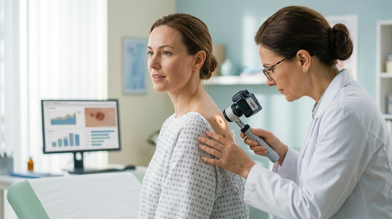
What Happens During Mole Mapping?
If mole mapping is part of your visit, it usually includes:
1. Full-body photography
- You’ll be asked to stand in specific positions while a camera captures standardized views of your entire body (front, back, sides, arms, legs, etc.).
- These images are stored securely and are used only for your medical care.
2. Close-up and dermoscopic images
- Any atypical moles or concerning spots are photographed more closely.
- A dermatoscope attached to a camera may be used to capture magnified details of the pigment patterns, blood vessels, and structure of the lesion.
3. Digital comparison over time
- At future visits, your dermatologist will compare new images to your baseline photos.
- They can quickly see:
- Whether new moles have appeared
- Whether existing moles have changed shape, color, or size
- Whether any lesions that seemed harmless before now look suspicious
This long-term record is particularly valuable for people with numerous or atypical moles.
If a Suspicious Spot Is Found
Not every suspicious-looking mole is cancer, and not every biopsy means cancer. But your doctor may recommend further steps:
Skin biopsy
- A small sample of skin is removed under local anesthesia (you are awake, but the area is numbed).
- Types of biopsy include shave, punch, or excisional biopsies, depending on the size and depth of the lesion.
- The sample is sent to a pathology lab to be examined under a microscope.
Waiting for results
- Results usually come back in days to a couple of weeks, depending on the clinic.
- The report will typically say whether the lesion is:
- Benign (non-cancerous)
- Precancerous (e.g., actinic keratosis)
- Cancerous (e.g., basal cell carcinoma, squamous cell carcinoma, or melanoma)
- If cancer is found, your dermatologist will discuss treatment options, which may include further surgery or, rarely, other therapies.
Remember: early detection usually means simpler treatment and better outcomes.
How Often Should You Get Screened?
The ideal frequency depends on your risk level and doctor’s advice, but some general patterns are:
- Average risk, no concerning factors: every 1–2 years or as recommended by your dermatologist.
- Higher risk (many moles, family history, fair skin, significant sun exposure): often every 6–12 months.
- Previous skin cancer or pre-cancerous lesions: your dermatologist may suggest more frequent follow-up, especially in the first few years after treatment.
Along with professional screenings, regular self-skin checks at home (once a month or every few months) help you notice changes early.
Tips for At-Home Skin Checks
Professional screening is best, but you’re still your own first line of defense. When examining your skin at home:
- Use a full-length mirror and a handheld mirror for hard-to-see areas.
- Check:
- Face, lips, and behind the ears
- Scalp (using a comb or asking a family member)
- Neck, chest, abdomen
- Under breasts
- Arms, hands, fingers, nails
- Back, buttocks, and back of legs
- Feet, including soles and between toes
- Look for:
- New moles or spots
- Moles that change in size, shape, or color
- Sores that don’t heal
- Any spot that is itchy, bleeding, crusted, or painful
If something concerns you, don’t wait—book a dermatologist appointment.
Key Takeaways
- Full-body skin cancer screenings are thorough head-to-toe exams designed to catch suspicious lesions early.
- Mole mapping creates a photographic record of your skin, allowing doctors to track changes over time and detect subtle signs of melanoma.
- People with higher risk—many moles, fair skin, strong sun exposure, or a family/personal history of skin cancer—benefit the most from regular screenings and mole mapping.
- Preparing well (clean skin, list of concerns, medical history) makes your appointment more efficient and effective.
- Finding a suspicious lesion is not a reason to panic; it’s an opportunity to treat problems early, when they’re most curable.
A practical next step is to schedule a visit with a dermatologist, especially if you’ve never had a full-body exam or if you’ve noticed a mole or spot that worries you. Early attention can make all the difference.

Leave a comment
This site is protected by hCaptcha and the hCaptcha Privacy Policy and Terms of Service apply.