
Article: Can You Diagnose Skin Conditions From a Photo? What You Need to Know
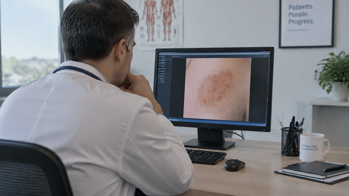
Can You Diagnose Skin Conditions From a Photo? What You Need to Know
It happens to almost everyone at some point. You notice a spot on your arm, a patch on your scalp, a discoloration on your leg, or a rash that appeared overnight. You do what millions of people do now: you pull out your phone, take a picture, and start searching. Within minutes you have typed the image's description into a search engine, uploaded it to an AI tool, or posted it in a forum asking strangers what they think. By the time you put the phone down, you either feel reassured or terrified, and neither feeling is reliably connected to what is actually happening on your skin.
So can you diagnose a skin condition from a photo? The honest answer is: sometimes, partially, and never with the certainty that a proper diagnosis requires. Photos are genuinely useful, and the technology behind them is improving fast. But there is a gap between "this image looks like something" and "this is what you have and here is how to treat it," and that gap is where people get hurt. Understanding exactly where the line falls will help you use photos wisely instead of either dismissing them entirely or trusting them too much.
What a photo actually captures
A photograph is a flat, two-dimensional record of reflected light. Dermatology, by contrast, is a three-dimensional, multisensory examination. When a dermatologist evaluates a lesion, they are gathering information that a picture simply cannot carry.
They look at the surface texture — whether it is smooth, scaly, crusted, or waxy. They touch it, feeling for firmness, mobility, and depth. They check whether it blanches under pressure, whether it is warm, whether it is tender. They use a dermatoscope, a specialized magnifying instrument with polarized light, to see structures beneath the surface that are invisible to a phone camera. They examine the rest of your body for similar lesions, because a single spot often means something different when it is accompanied by twenty others. They ask about your history: how long it has been there, whether it itches or hurts, whether it bleeds, whether it changes. They look at your mucous membranes, your nails, your scalp, your lymph nodes. They ask what medications you take and what you do for work.
A photo of one spot on your forearm carries almost none of that. It gives you color, shape, border, and rough texture, and it flattens even those. Color is notoriously distorted by lighting, white balance settings, and skin tone rendering in phone cameras. Scale is lost without a reference object. Depth is invisible. The surrounding skin that might show a pattern is cropped out.
This is not an argument that photos are useless. It is an argument that photos solve a different problem than the one people usually think they are solving.
Where photos genuinely help
There are real, well-established uses for skin photography, and it is worth being clear about them because they are not trivial.
Tracking change over time. A single photo is one data point. A series of photos taken in consistent lighting, from the same angle, with the same camera and a ruler or coin in frame, is a legitimate record. Dermatologists frequently recommend this for people with many moles or a history of skin cancer. If a lesion is growing, changing color, or developing irregular borders, a dated photo series makes that visible in a way memory cannot. Memory is unreliable; people routinely cannot say whether a mole looked different six months ago.
Communicating with a clinician. If you have an established relationship with a dermatologist and a lesion you are worried about, sending a photo ahead of a visit can help them triage. Many health systems now run store-and-forward teledermatology programs where a patient submits images and a dermatologist reviews them asynchronously. These programs work because a trained professional is interpreting the image within a full clinical context — your history, your other medications, your risk factors, and the option to bring you in if the picture is ambiguous.
Documenting for yourself. If you have a chronic condition like psoriasis, eczema, or acne, photos help you see whether a treatment is working. Again, consistent framing and lighting matter enormously. A photo taken in bright bathroom light and one taken in a dim bedroom the next week are not comparable.
Raising the question. A photo can prompt you to seek care. That is a real benefit. Someone who notices an odd lesion while taking a selfie and then calls a clinic has used the camera well. The camera started a process; it did not finish one.
Where photos fail
Now the harder part. Here is what goes wrong when people treat a photo as a diagnosis.
Many conditions look alike. This is the central problem in dermatology and it does not go away with better cameras. Eczema, psoriasis, ringworm, contact dermatitis, pityriasis rosea, and dozens of other conditions can produce red, scaly, itchy patches that are genuinely difficult to tell apart from an image alone. Melanoma can resemble a benign mole. A basal cell carcinoma can look like a pimple that will not heal. Seborrheic keratoses, which are harmless, are notorious for looking like melanomas and vice versa. Two different conditions can even coexist on the same patch of skin.
Rare things look like common things. Search engines and image-matching tools are trained on what is most common, which means common conditions dominate the results. But when you upload a photo, you are usually looking for reassurance about the worst possibility, not the most likely one. The reverse mismatch — a rare serious condition that superficially resembles a common benign one — is exactly the scenario where a photo-based guess is most dangerous.
Lighting and skin tone distort everything. Images taken under warm indoor bulbs shift reds and yellows. Fluorescent light shifts greens. Phone cameras apply automatic color correction that varies by device and by the scene. Conditions that appear as subtle color changes can wash out entirely. This problem is worse for people with darker skin tones, where erythema — the redness that signals inflammation — presents differently and is often poorly captured by consumer cameras and, historically, poorly represented in the training data behind diagnostic tools.
Context is missing. Whether a lesion is new or old, whether it is one of many, whether it appeared after starting a new medication, whether there is a family history of skin cancer, whether you have been in the sun, whether you have diabetes or an autoimmune condition — none of this is in the photo. The same image can mean "watch it" or "come in today" depending entirely on that missing information.
The stakes are asymmetric. If you look at a photo of a rash and decide it is nothing, and you are wrong, you may lose weeks or months. In the case of melanoma, that delay can be the difference between an easy excision and a serious prognosis. The cost of a wrong "it's fine" is much higher than the cost of a wrong "get it checked."
What about AI and diagnostic apps?
This is the part of the conversation that has changed most in recent years, and it deserves a careful answer rather than a blanket dismissal.
There is real research on machine learning for dermatology, and some of it is genuinely impressive. Studies have shown that convolutional neural networks can classify certain skin lesions — particularly dermoscopic images of pigmented lesions — with accuracy comparable to dermatologists in controlled settings. Some regulatory bodies have cleared AI-assisted tools for specific, narrow uses, typically as a second reader for dermoscopic images of pigmented lesions in a clinical workflow, with a clinician in the loop.
Notice the qualifiers in that paragraph, because they carry all the weight. Narrow use. Dermoscopic images, not phone snapshots. Pigmented lesions specifically, not "skin conditions" generally. In a clinical workflow. With a clinician reviewing.
Consumer apps that claim to analyze a phone photo and tell you what you have are operating in a very different regime. The image quality is uncontrolled. The conditions are not limited to a well-characterized set. There is no clinical history. And the output is often presented with a confidence that the underlying evidence does not support. Some of these apps have been studied, and the results have been mixed at best, with performance dropping sharply on real-world phone images compared to the curated datasets used in development.
Search engines that match your photo to similar images have an additional problem: they are matching appearance, not pathology. They will return the most visually similar images in their index, which may have nothing to do with what you actually have. A rare condition with a distinctive look may outrank a common one with a subtler presentation.
None of this means the technology is worthless or that it will never get better. It means that in 2026, the responsible framing is that these tools can suggest possibilities, not deliver diagnoses, and that any tool claiming otherwise is overclaiming.
The specific danger of reassurance
There is a psychological trap worth naming directly, because it is where most photo-based harm actually occurs.
When you are worried about a spot, you want an answer. A photo tool gives you an answer instantly, and the answer you are hoping for is "it's nothing." If the tool says "this appears benign," you feel relief, and that relief makes it much harder to override the result with common sense. The asymmetry of the situation — a low-effort action producing a high-value emotional payoff — is almost perfectly designed to discourage you from seeking real care.
The reverse also happens. Someone uploads a photo of a completely ordinary mole, the tool returns a scary-sounding list of possibilities, and they spend a week terrified. Both failure modes come from the same source: treating a probabilistic guess about appearance as a conclusion about your health.
What you should actually do
If you have a skin concern, here is a practical framework that uses photos well without relying on them.
Photograph it for the record. Take a clear photo in natural daylight, with the camera perpendicular to the skin, with a ruler, coin, or tape measure in frame for scale. Note the date. If you have multiple lesions, photograph each one and note its location. This takes two minutes and gives a dermatologist useful information.
Apply the change criteria. Regardless of what any app or search tells you, these findings warrant a professional evaluation: a lesion that is new and changing; one that has grown in size, changed shape, or changed color; one with irregular, notched, or blurred borders; one that is larger than a pencil eraser and new; one that bleeds, itches, crusts, or does not heal within a few weeks; anything that hurts; anything that appears on a mucous membrane; a mole that stands out from the others on your body. These are the widely used warning signs, and they do not require a photo to evaluate. They require a clinician.
Know your personal risk. A history of sunburn, use of tanning beds, a family history of melanoma, a large number of moles, a suppressed immune system, or certain genetic conditions raise your baseline risk substantially. If any apply to you, your threshold for getting something checked should be lower.
Use photos as a bridge, not a destination. Send the photo to your clinician if your health system supports it. Bring the photo to your appointment. Use it to ask a specific question. Do not use it as a substitute for the appointment.
Be skeptical of confident claims. If an app or website tells you with certainty what you have based on a photo, that certainty is not earned. Real diagnosis involves uncertainty, and a good clinician will tell you when they are uncertain and what they plan to do about it.
What a real diagnosis looks like
For contrast, here is roughly what happens when a dermatologist evaluates a concerning lesion. They take a history. They examine the lesion and the surrounding skin, often with a dermatoscope. They may examine your entire body. They may compare the lesion to your other moles. They may photograph it themselves for the record. If it looks suspicious, they may perform a biopsy — removing a small sample and sending it to a pathologist, who examines it under a microscope and issues a report. In some cases, the diagnosis is not confirmed until that pathology report comes back. That is the standard. Nothing you can do with a phone photo approaches it, and that is not a limitation of the camera so much as a description of what diagnosis actually is.
A note on where you are
Access to dermatology varies enormously by location, and this shapes how people actually use photo tools. In places with long wait times or few dermatologists per capita, the temptation to self-diagnose from a photo is higher, and the consequences of a missed diagnosis are worse. Telehealth has improved this considerably, and store-and-forward dermatology services have made specialist input available in places that previously had none. If that is your situation, the right move is to find a telehealth service with real clinicians rather than to rely on an app. In the Philippines and across much of Southeast Asia, teledermatology via established hospital systems and telehealth platforms is increasingly available, and it is a far better use of your phone than any diagnostic app.
The bottom line
Can you diagnose skin conditions from a photo? No, not reliably, and the failure modes are not random — they cluster around exactly the cases where getting it wrong matters most. Photos are excellent for tracking change over time, for communicating with a clinician, and for prompting you to seek care. They are poor substitutes for examination, history, and, when needed, a biopsy.
The best use of your phone camera is as the beginning of a conversation with someone qualified, not as a replacement for it. Take the photo. Note the date. Apply the change criteria. And if something is new, changing, bleeding, or simply worrying you, put the phone down and call someone who can actually look at it.
This article is general information, not medical advice. If you have a skin concern, consult a qualified healthcare professional.

Leave a comment
This site is protected by hCaptcha and the hCaptcha Privacy Policy and Terms of Service apply.