
Article: Can You Diagnose Skin Conditions From a Photo? What You Need to Know
Can You Diagnose Skin Conditions From a Photo? What You Need to Know
It's 11 p.m., you're lying in bed, and you've just noticed a mole on your shoulder that you swear wasn't there last year. Or a patch of dry, scaly skin on your elbow that's been itching for a week. Within thirty seconds you can have a photo of it uploaded to an app, an AI tool, or a "spot check" service, and within a minute you can have an answer.
Sometimes that answer is reassuring. Sometimes it's terrifying. And sometimes it's flat-out wrong.
So the question is simple and worth answering carefully: can you actually diagnose a skin condition from a photo? The honest answer is that photos are genuinely useful for some things, unreliable for others, and dangerous when treated as a final verdict. Here's what the evidence says, where photo-based assessment helps, where it fails, and how to use it without gambling with your health.
The Short Answer
No, a photo alone cannot reliably diagnose most skin conditions, but it can be a legitimate first step — especially when the image is reviewed by a qualified clinician rather than interpreted by an algorithm alone.
That distinction matters more than most people realize. "Diagnosing from a photo" describes three very different things:
- A doctor reviewing a photo you sent them as part of an existing care relationship
- A telehealth service where a dermatologist reviews images and your history
- A consumer app or AI tool that analyzes an image and returns a label or risk score
These three carry very different levels of reliability, and lumping them together is where people get into trouble.
Why Skin Is Uniquely Suited to Pictures — and Uniquely Tricky
Dermatology has always been a visual specialty. Dermatologists are trained to recognize patterns: the shape, border, color, symmetry, and surface of a lesion. That's why images have real clinical value. A well-lit, in-focus photo of a rash can genuinely help a doctor who knows your history narrow down possibilities.
But skin has characteristics that make image-only assessment harder than it looks:
Depth is invisible in a photo. A raised nodule, a shallow ulcer, and a flat pigmented patch can look similar in a two-dimensional image, but they mean completely different things. Texture and induration — the firmness of tissue when pressed — carry diagnostic weight that a photograph simply cannot transmit.
Color is unreliable. Phone cameras apply automatic white balance, saturation, and color correction. The same mole photographed under a warm bedside lamp, in daylight, and with flash can appear three different colors. In dermatology, color is not a minor detail; it's a primary diagnostic criterion. Overcorrected images can make a benign lesion look darker, or wash out the subtle variation that signals trouble.
Scale and location are ambiguous. A lesion that looks alarming at macro distance might be a tiny, harmless freckle. Without a ruler, a reference object, or a clear anatomical landmark, the image can mislead.
The rest of the body is missing. Many skin conditions — psoriasis, eczema, drug reactions, scabies, contact dermatitis — are diagnosed by their distribution across the body. A photo of one arm cannot show that the same rash appears on both elbows and both knees, which is exactly the pattern that points to psoriasis.
Your history is missing. When did it appear? Does it itch, burn, or bleed? Have you started a new medication? Is there a family history of skin cancer? What's your occupation? These questions often matter more than the image itself. Diagnosing from a picture with no context is like reading one sentence from the middle of a novel.
Some of the most dangerous conditions look boring. Early melanoma can look like an ordinary mole. Early basal cell carcinoma can look like a pimple that won't heal. Amelanotic melanoma has little or no pigment at all, which makes it especially easy to miss — by eye, by camera, and by algorithm.
What Photo-Based Assessment Is Actually Good At
This isn't a story about technology being useless. Image-based skin assessment has legitimate, well-documented applications.
Triage and prioritization. A photo can help a clinician decide how urgently you need to be seen. "This needs a biopsy this week" versus "this can wait for a routine appointment" is a meaningful decision that an image can support.
Monitoring over time. This is arguably the strongest use case. A consistent photo taken with the same lighting, angle, and distance can reveal change — a lesion growing, changing shape, developing irregular borders. Change over time is one of the most important signals in dermatology, and photos are excellent at capturing it.
Teledermatology in access-limited settings. In places where dermatologists are scarce or travel is difficult, store-and-forward teledermatology has expanded access to specialist opinions. Studies generally show good concordance between teledermatology assessment and in-person diagnosis for many common conditions — but with important caveats about which conditions and how the images were captured.
Follow-up after treatment. If you've started a treatment for acne, eczema, or a fungal infection, photos can help track whether it's working.
Documenting for your own records. Even if no one diagnoses from your photo, having a dated, consistent record can be genuinely helpful when you finally see a clinician.
Where Photo-Based Diagnosis Fails
Now the part that matters most.
AI skin apps have a specificity problem. Research on consumer-facing dermatology apps and AI classifiers has repeatedly found that while sensitivity — the ability to flag something suspicious — can look impressive in headlines, the tools often produce high false-positive rates. That means a lot of benign lesions get flagged as potentially malignant, and a lot of different conditions get collapsed into the same few labels. A study of popular skin cancer detection apps found that many performed poorly compared to clinicians, with some apps misclassifying a substantial share of images.
Algorithms are trained on biased data. If a model was trained mostly on images of lighter skin, which is common, its accuracy on darker skin tones can drop significantly. This is a known and documented equity problem in dermatology AI. If you have darker skin, you should be especially skeptical of a consumer app's assessment.
Rare conditions are invisible to models. An AI trained on common conditions may have almost no examples of unusual presentations. Rather than saying "I don't know," many tools will force the image into the closest category they recognize.
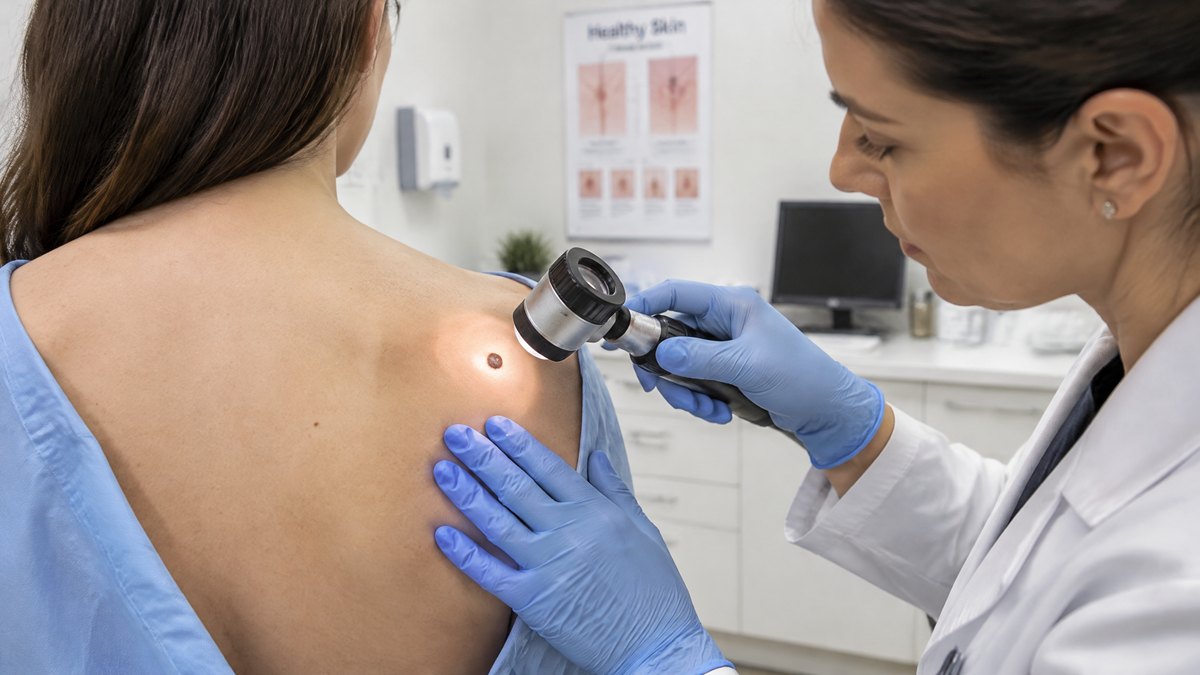
Photos can miss what eyes catch. In-person examination includes palpation, dermoscopy, and a full-body scan — looking for lesions the patient didn't notice or photograph. A patient-directed photo only covers what the patient already found. That's a fundamental blind spot no algorithm can fix.
The stakes are asymmetric. A false negative — telling someone a melanoma is "probably benign" — can cost months of delay in a disease where early detection is everything. A false positive causes anxiety, unnecessary biopsies, and cost. Neither error is acceptable, and no consumer photo tool has proven it avoids both.
Regulatory status is murky. Many consumer skin apps are not cleared as medical devices and are not intended to diagnose anything. Their terms of service frequently say exactly that, buried in language nobody reads. A tool that says "this looks like it could be eczema" is not a diagnosis, even when it's phrased like one.
The Role of Teledermatology Done Properly
There's a crucial distinction between an unregulated app and a real teledermatology consultation.
In a proper teledermatology visit:
- A licensed clinician reviews your images, not a standalone algorithm
- You provide a history: onset, symptoms, medications, family history, prior treatments
- The clinician can ask follow-up questions or request additional images
- The clinician can order a biopsy or refer you for in-person examination
- There is a documented plan and someone accountable for the outcome
In this context, photos are a tool within a clinical process. That's fundamentally different from an app returning a percentage risk score with no clinician in the loop.
Even here, teledermatology has limitations: it may be inappropriate for lesions that need palpation or dermoscopy, and most reputable services will tell you when you need to be seen in person.
How to Take a Photo That's Actually Useful

If you're going to send an image to a clinician — or keep one for tracking — quality matters enormously. A poor photo can be worse than no photo, because it can mislead.
Use good, indirect daylight. Stand near a window. Avoid direct flash, which creates glare and color cast, and avoid warm indoor bulbs, which shift colors orange.
Include a size reference. A coin, a ruler, or a tape measure next to the lesion gives scale.
Take multiple angles and distances. One wide shot showing the body location with surrounding anatomy, one close-up filling the frame with the lesion, and one at an oblique angle to show whether it's raised.
Don't zoom digitally. Move the camera closer instead. Digital zoom destroys detail.
Use a plain background if possible. Skin against skin is fine; a busy patterned background makes it harder to judge color and border.
Keep lighting consistent for tracking. If you're monitoring over time, take every photo in the same spot, same light, same distance, ideally same time of day.
Photograph the whole area, not just the dot. Include a margin of normal skin around the lesion so the border is visible.
Take photos of everything you're worried about. If a clinician is reviewing remotely, they need the full picture — literally.
Warning Signs That Mean: Don't Rely on a Photo
Some findings should send you to an in-person appointment regardless of what any app says.
The classic melanoma checklist uses ABCDE:
- A — Asymmetry: one half doesn't match the other
- B — Border: irregular, ragged, blurred, or notched edges
- C — Color: multiple colors within one lesion, or uneven distribution
- D — Diameter: larger than about 6 mm (a pencil eraser), though smaller melanomas exist
- E — Evolving: any change in size, shape, color, elevation, or new symptoms like itching or bleeding
There's also the "ugly duckling" sign: if one mole looks different from all the others on your body, it deserves attention even if it doesn't tick the ABCDE boxes.
Beyond moles, see someone in person if you have:
- A sore or pimple that hasn't healed in three to four weeks
- A lesion that bleeds, crusts, or oozes without obvious cause
- Rapidly spreading redness, especially with fever or pain
- A rash covering a large portion of your body
- Blistering, peeling, or involvement of the eyes, mouth, or genitals
- Any skin change accompanied by systemic symptoms — fever, joint pain, weight loss, fatigue
- Severe pain or swelling around a skin lesion
These presentations demand physical examination, and often bloodwork, cultures, or biopsy.
A Practical Framework for Using Photo Tools Wisely
If you want to use photo-based tools without putting yourself at risk, here's a reasonable approach:
Use them for questions, not answers. A photo tool can help you formulate "should I be worried about this?" and prompt you to seek care. It should never be the reason you don't seek care.
Treat a reassuring result as uninformative. A tool saying "likely benign" tells you almost nothing. It doesn't clear you.
Treat a suspicious result as a signal to act. If a tool flags something, that's a reasonable trigger to book an appointment — not to panic, but to get a real assessment.
Never let an app override your instincts. If something feels wrong, is changing, or is bothering you, that alone justifies a visit. Patients are often the first to notice their own melanoma.
Check who's actually reading your image. Is there a licensed clinician? Where are they licensed? Is there a follow-up pathway? If not, you're using an entertainment product with medical-sounding output.
Prioritize in-person care when it's available. If you can see a dermatologist, see a dermatologist. A photo is a substitute for access, not for expertise.
The Bottom Line
Can you diagnose skin conditions from a photo? Not reliably — and anyone promising otherwise is overselling.
What photos can do is help a qualified clinician assess, triage, and monitor your skin as part of a real clinical process. What they cannot do is replace physical examination, history-taking, dermoscopy, biopsy, and clinical judgment. Consumer AI apps, however sophisticated their marketing, remain unproven as diagnostic tools and carry documented risks around false positives, false negatives, and biased performance across skin tones.
The most useful way to think about a skin photo is this: it's a question you're asking, not an answer you're receiving. Use it to start a conversation with someone qualified. Use it to track change over time so that conversation is better informed. But never let a picture, an algorithm, or a percentage score make a decision that belongs to a clinician and to you.
If something on your skin worries you, the photo is step one. The appointment is step two. Don't stop in the middle.

Leave a comment
This site is protected by hCaptcha and the hCaptcha Privacy Policy and Terms of Service apply.