
Understanding Lentigo Maligna: Is That Facial Sun Spot Dangerous?
A new brown patch on the face can be easy to dismiss as “just sun damage”. After all, many people develop freckles, age spots and uneven pigmentation as they get older, especially on areas that have seen years of sunlight. But some facial sun spots deserve closer attention. One of them is lentigo maligna, a slow-growing form of melanoma that often appears on sun-damaged skin.
Lentigo maligna can look deceptively harmless at first. It may start as a flat, brown mark on the cheek, nose, temple, forehead or ear. It might grow so gradually that months or even years pass before anyone becomes concerned. Yet despite its slow pace, lentigo maligna matters because it is a type of melanoma in its earliest stage. If it remains confined to the top layer of the skin, it is highly treatable. If it grows deeper, it can become lentigo maligna melanoma, which has the potential to spread.
Understanding what lentigo maligna looks like, who is most at risk and when to seek medical advice can make a real difference. Not every facial sun spot is dangerous, but any changing or unusual patch should be checked by a qualified health professional.
What is lentigo maligna?
Lentigo maligna is a type of melanoma in situ. “In situ” means the abnormal cells are still limited to the outermost layer of the skin, the epidermis. At this stage, the melanoma cells have not invaded deeper tissues. That is why early diagnosis and treatment are so important.
It most often develops on chronically sun-exposed skin, particularly the face and neck. Unlike some other melanomas that may appear suddenly or grow quickly, lentigo maligna usually develops slowly. It is especially associated with long-term, cumulative exposure to ultraviolet radiation rather than a single episode of sunburn.
The condition is more common in older adults, particularly those with fair skin, a history of significant sun exposure, and visible sun damage. However, it can occur in anyone. People with darker skin tones are less commonly affected, but melanoma can still develop and may be diagnosed later if changes are harder to spot or are not recognised quickly.
How is it different from an ordinary sun spot?
A typical sun spot, sometimes called a solar lentigo or age spot, is a flat area of increased pigmentation caused by sun exposure. These spots are usually harmless. They tend to be evenly coloured, clearly defined and fairly stable over time.
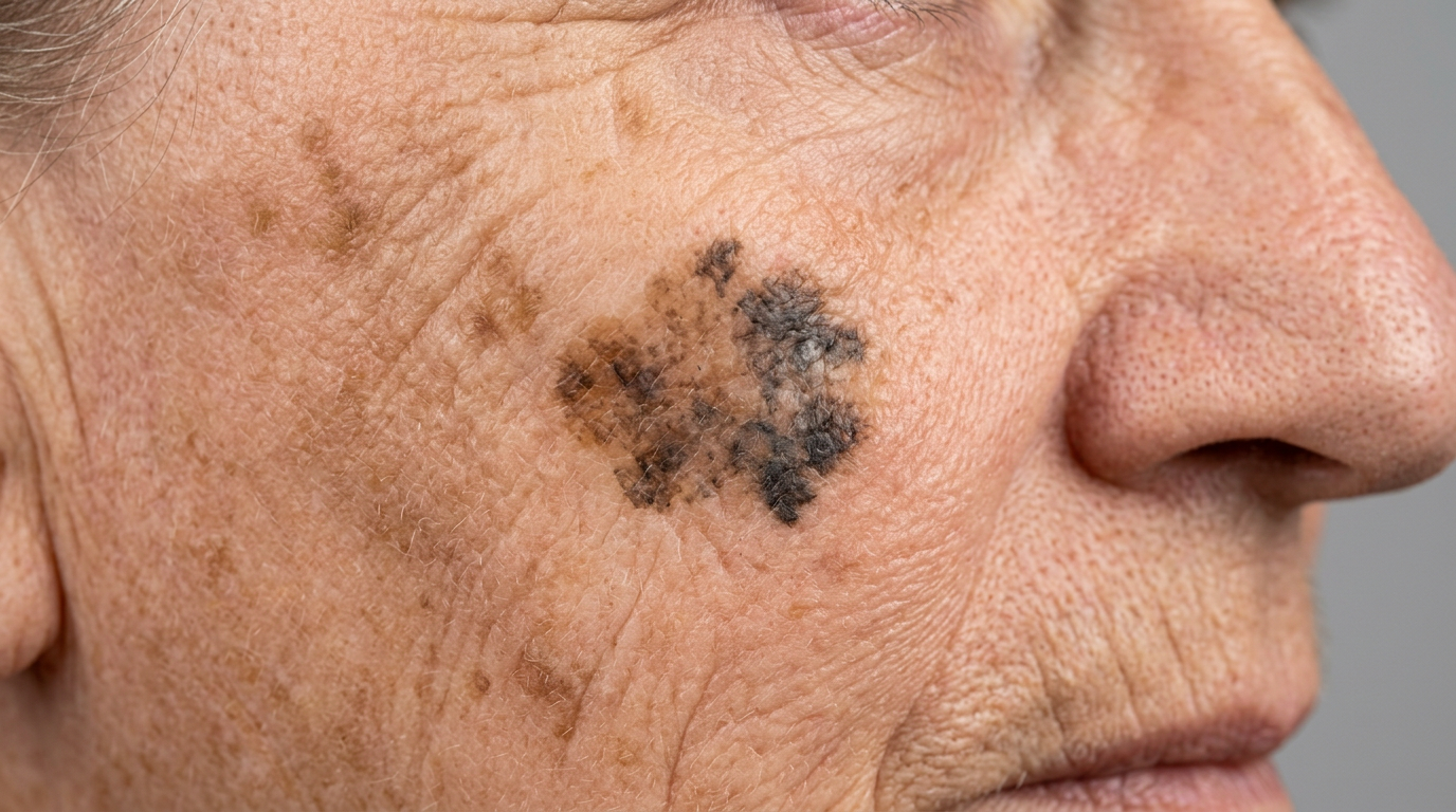
Lentigo maligna can resemble an ordinary sun spot, but there are warning signs that set it apart. It may have an irregular outline, several shades of brown or black, or areas that look grey, pink, white or bluish. It may slowly expand, darken or become patchier. The edges may look blurred rather than crisp.
The difficulty is that these differences can be subtle. A harmless sun spot and early lentigo maligna may look similar to the untrained eye. This is one reason dermatologists often use a dermatoscope, a handheld magnifying device with special lighting, to examine suspicious pigmented lesions more closely.
What does lentigo maligna look like?
Lentigo maligna often appears as a flat, unevenly pigmented patch on sun-damaged skin. It may be tan, light brown, dark brown or black, and it may contain more than one colour. The shape is often irregular rather than round or oval.
Common features include:
- A slowly enlarging patch, often present for months or years.
- Uneven colour, with different shades within the same mark.
- Irregular or notched borders, rather than smooth edges.
- A size larger than many ordinary freckles, sometimes several centimetres across.
- A location on the face, especially the cheeks, temples, nose, forehead, ears or scalp.
Some lesions are quite faint, particularly in the early stages. Others may look like a smudge of brown pigment, a large freckle, or a patch of uneven sun damage. Occasionally, part of the lesion becomes raised, thicker, darker or develops a lump. That can be a sign that it has started to invade more deeply and needs urgent assessment.
Why does lentigo maligna develop?
The main cause is long-term damage from ultraviolet radiation, usually from the sun. Over time, UV radiation can damage DNA in skin cells. If that damage affects the cells that produce pigment, known as melanocytes, abnormal growth can occur.
Lentigo maligna is linked more strongly with chronic sun exposure than with intermittent intense sunburn. This is why it often appears on areas that have been exposed every day for decades: the face, neck, ears, scalp and backs of the hands.
Risk factors include:
- Older age, because UV damage accumulates over time.
- Fair skin, especially skin that burns easily.
- A history of heavy sun exposure, including outdoor work or frequent outdoor recreation.
- Previous skin cancers, including melanoma or non-melanoma skin cancers.
- Many freckles or visible sun damage, such as rough patches, wrinkles and pigmentation.
- A weakened immune system, which can reduce the body’s ability to control abnormal cells.
- Use of tanning beds, which expose the skin to harmful UV radiation.
Having one or more risk factors does not mean someone will definitely develop lentigo maligna. Equally, having no obvious risk factors does not make a changing facial patch safe to ignore.
Is lentigo maligna dangerous?

Lentigo maligna is serious because it is a form of melanoma, but it is usually very treatable when diagnosed early. While it remains “in situ”, the abnormal cells are confined to the surface layer of the skin. At that stage, treatment aims to remove or destroy the abnormal cells before they become invasive.
The danger increases if lentigo maligna develops into lentigo maligna melanoma. This means the melanoma cells have grown deeper into the skin. Once melanoma becomes invasive, there is a risk, depending on depth and other features, that it can spread to lymph nodes or other parts of the body.
The reassuring point is that lentigo maligna often grows slowly, giving an opportunity for early detection. The less reassuring point is that its slow growth can lead people to delay seeking help. A patch that has been “there for years” can still be important, especially if it has changed.
When should you get a facial sun spot checked?
Any new, changing or unusual pigmented mark on the face should be assessed, particularly if it looks different from your other marks. Dermatologists often refer to the ABCDE warning signs of melanoma:
- Asymmetry: one half does not match the other.
- Border: the edges are irregular, blurred or notched.
- Colour: there are multiple colours or uneven shading.
- Diameter: the mark is larger than about 6 mm, although melanomas can be smaller.
- Evolving: the mark is changing in size, shape, colour, texture or symptoms.
For lentigo maligna, the “E” for evolving is especially important. Because it may grow slowly, changes can be hard to notice from day to day. Comparing photographs over several months can sometimes reveal enlargement or colour variation that was not obvious.
You should seek prompt medical advice if a facial spot:
- Is getting bigger, even gradually.
- Has several colours or is becoming darker in places.
- Has an irregular shape or uneven border.
- Starts to itch, bleed, crust or become tender.
- Develops a raised area or lump within a previously flat patch.
- Looks unlike your other freckles or sun spots.
It is better to have a harmless mark checked than to overlook an early melanoma.
How is lentigo maligna diagnosed?
A doctor or dermatologist will usually begin by examining the skin and asking how long the mark has been present, whether it has changed, and whether there is any history of skin cancer or significant sun exposure.
The lesion may be examined with a dermatoscope. This can reveal patterns and structures that are not visible to the naked eye. If lentigo maligna is suspected, a skin biopsy is usually needed. A biopsy involves removing a small sample, or sometimes the whole lesion, so it can be examined under a microscope.
Diagnosis can be more complex on the face because sun-damaged skin may contain widespread pigmentation, and the edges of lentigo maligna can be difficult to define. In some cases, multiple samples are needed to map the affected area accurately.
How is it treated?
Treatment depends on the size, location, biopsy findings and the person’s overall health. The aim is to remove or control all abnormal cells while preserving as much healthy tissue and function as possible, particularly on cosmetically sensitive areas of the face.
The most common treatment is surgical removal. This may involve standard excision, where the lesion is removed with a margin of normal-looking skin around it. In some cases, staged excision or Mohs surgery may be used. These techniques allow careful checking of the edges to make sure the abnormal cells have been fully removed, which can be especially useful on the face.
Other treatments may be considered when surgery is not suitable. These can include radiotherapy or topical medicines in selected cases, but they are not appropriate for everyone and require careful specialist guidance. Because lentigo maligna can have hidden extensions beyond what is visible on the surface, treatment choice should be individualised.
After treatment, follow-up is important. People who have had lentigo maligna may be at increased risk of developing another skin cancer, so regular skin checks and good sun protection are essential.
Can lentigo maligna be prevented?
Not all cases can be prevented, but reducing UV exposure lowers the risk of further sun damage and skin cancers. Prevention is particularly important for people with fair skin, a history of skin cancer or extensive sun damage.
Practical sun protection includes:
- Wearing a broad-spectrum sunscreen with high SPF on exposed skin every day.
- Reapplying sunscreen during prolonged outdoor activity, sweating or swimming.
- Using a wide-brimmed hat to protect the face, ears and neck.
- Wearing UV-protective sunglasses and protective clothing.
- Seeking shade, especially during periods of strong sunlight.
- Avoiding tanning beds, which increase UV damage.
- Checking the skin regularly for new or changing marks.
Daily incidental exposure matters. Many people remember sunscreen on holidays but forget it during routine activities such as walking, gardening, driving or sitting near a sunny window. Over years, those small exposures add up.
How to monitor a suspicious sun spot
If you have a facial mark that you are watching, take clear photographs in good lighting. Include a ruler or another size reference if possible. Repeat the photo every month or two and compare images side by side. Do not rely on memory alone, as gradual changes are easy to miss.
That said, monitoring is not a substitute for medical assessment. If a mark already has concerning features, book an appointment rather than waiting to see what happens. A dermatologist can decide whether the lesion needs biopsy, photography, monitoring or treatment.
Avoid trying to diagnose the mark yourself using online images. Lentigo maligna varies widely in appearance, and many harmless marks can look worrying in photographs. The reverse is also true: an early melanoma may appear quite subtle.
The bottom line
Most facial sun spots are harmless, but some are not. Lentigo maligna is a slow-growing form of melanoma that often appears as a flat, irregular brown patch on sun-damaged skin of the face. It may look like an age spot at first, but warning signs include gradual enlargement, uneven colour, irregular borders, and any new raised, bleeding or crusted area.
The key message is simple: do not panic, but do not ignore change. Lentigo maligna is usually highly treatable when found early, before it becomes invasive. If a facial sun spot is new, changing, irregular or simply does not look right, arrange a professional skin check. Early attention can turn a potentially dangerous mark into a manageable diagnosis.



Leave a comment
This site is protected by hCaptcha and the hCaptcha Privacy Policy and Terms of Service apply.