Article: Mole Screening: What You Need to Know to Protect Your Skin
Mole Screening: What You Need to Know to Protect Your Skin
Every person on Earth has moles. Most adults carry somewhere between 10 and 40 of them — small clusters of pigmented cells scattered across the body like a constellation unique to each individual. The vast majority of these marks are completely harmless, quiet passengers on the skin that never cause a moment of trouble. But in a small number of cases, a mole can signal something far more serious: melanoma, one of the most aggressive forms of skin cancer.
That's where mole screening comes in — and why understanding it could, without exaggeration, save your life.
What Is Mole Screening?
Mole screening is a medical examination designed to evaluate the moles and pigmented lesions on your skin for signs of abnormality. It can be performed by a dermatologist, a trained nurse, or even initiated by you at home through regular self-checks.
The purpose is straightforward: catch dangerous changes early, before a potentially cancerous mole has the chance to grow, spread, or become life-threatening. Early-stage melanoma, when detected before it penetrates deeper layers of the skin, has a five-year survival rate of over 99%. Once it spreads to distant organs, that figure drops dramatically. The difference between those two outcomes often comes down to one thing — screening.
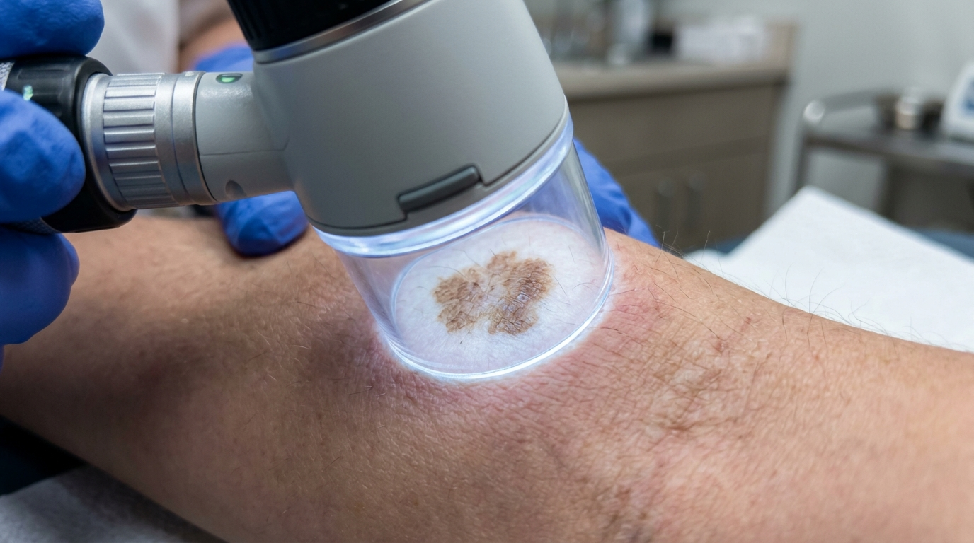
A professional mole screening typically involves a full-body skin examination. The dermatologist visually inspects the skin from scalp to soles, paying close attention to any moles that stand out in size, shape, color, or texture. In many clinics, a tool called a dermatoscope — a specialized magnifying instrument with polarized light — is used to examine suspicious moles at a level of detail invisible to the naked eye. Some practices also use digital mole mapping, a photographic technique that captures high-resolution images of the entire body surface, creating a baseline record that makes it easy to spot new or changing moles at future appointments.
Why Does Mole Screening Matter?
Skin cancer is the most common cancer worldwide, and melanoma accounts for the majority of skin cancer deaths despite being less common than basal cell or squamous cell carcinoma. The reason is its tendency to metastasize — to send cancerous cells through the lymphatic system and bloodstream to other parts of the body.
The challenge with melanoma is that it often begins in or near an existing mole, and in its earliest stages, the changes can be subtle. A slight darkening here, an uneven edge there — details easy to overlook during the rush of daily life. Professional screening exists precisely because trained eyes catch what untrained ones miss. Dermatologists are skilled at distinguishing between a benign mole that's simply evolved with age and one that's displaying the early warning signs of malignancy.
Beyond melanoma, screening can also detect other skin conditions worth monitoring: dysplastic nevi (atypical moles that carry a higher statistical risk of becoming cancerous), basal cell carcinomas, and various benign but cosmetically concerning growths. A screening appointment isn't just about cancer — it's a comprehensive check-in on the health of your largest organ.
Who Should Get Screened?
The short answer: everyone benefits from periodic skin checks. But certain groups carry higher risk and should prioritize regular professional screening:
- Fair-skinned individuals — People with lighter skin, light eyes, and red or blonde hair produce less melanin, the pigment that offers some natural UV protection. This demographic faces a significantly higher melanoma risk.
- Those with a high mole count — Having more than 50 common moles, or more than 5 atypical (dysplastic) moles, elevates risk.
- Family history of melanoma — A first-degree relative (parent, sibling, child) diagnosed with melanoma roughly doubles your own risk.
- Personal history of sunburns — Particularly blistering sunburns during childhood and adolescence, which cause lasting DNA damage to skin cells.
- Frequent sun exposure or tanning bed use — Cumulative UV radiation is the single largest modifiable risk factor for skin cancer.
- Immunosuppressed individuals — Organ transplant recipients, people on long-term immunosuppressive medications, and those with certain autoimmune conditions face elevated skin cancer rates.
Even if none of these risk factors apply to you, a baseline screening in your twenties or thirties gives your dermatologist a reference point for comparison in the years ahead. Think of it as establishing a map — one that makes any future deviation far easier to spot.
The ABCDEs of Self-Screening

Between professional appointments, monthly self-examinations are one of the simplest and most effective habits you can build. The widely taught ABCDE framework gives you a practical checklist for evaluating any mole on your body:
- A — Asymmetry. If you draw an imaginary line through the middle of the mole, do the two halves match? Asymmetry is a red flag.
- B — Border. Healthy moles tend to have smooth, well-defined edges. Irregular, scalloped, or blurred borders deserve a closer look.
- C — Color. A single, uniform shade of brown is typical. Multiple colors within one mole — brown mixed with black, red, white, or blue — can signal trouble.
- D — Diameter. Moles larger than 6 millimeters (roughly the size of a pencil eraser) warrant attention, though melanomas can certainly be smaller at the time of detection.
- E — Evolution. This is arguably the most important letter. Any mole that is changing — in size, shape, color, elevation, or symptoms like itching and bleeding — should be evaluated promptly.
To perform a thorough self-check, stand in front of a full-length mirror in a well-lit room. Use a hand mirror to inspect hard-to-see areas: the back of your neck, your scalp (part your hair methodically), between your toes, the soles of your feet, and behind your ears. Don't skip the areas that rarely see sunlight — melanoma can appear anywhere, including the palms, under fingernails, and on mucous membranes.
What Happens If Something Looks Suspicious?
If your dermatologist identifies a mole that raises concern during screening, the next step is usually a biopsy. This is a quick, minimally invasive procedure performed under local anesthesia. The doctor removes all or part of the mole and sends the tissue to a pathology lab for microscopic analysis.
Results typically come back within one to two weeks and fall into a few categories:
- Benign — The mole is non-cancerous. No further treatment needed.
- Dysplastic (atypical) — The mole shows unusual cellular features but isn't cancerous. Depending on the degree of atypia, your doctor may recommend removing the remaining tissue as a precaution and scheduling closer follow-up.
- Melanoma in situ — Cancerous cells are present but confined to the outermost layer of skin (the epidermis). Surgical excision with clear margins is typically curative.
- Invasive melanoma — The cancer has penetrated deeper into the skin. Treatment depends on the stage and may involve wider excision, sentinel lymph node biopsy, immunotherapy, targeted therapy, or radiation.
The critical takeaway is that a biopsy is not a diagnosis of cancer — it's a diagnostic tool. Most biopsied moles turn out to be benign. The minor discomfort of the procedure is an extraordinarily small price to pay for the certainty it provides.
Technology Is Changing the Game
The landscape of mole screening is evolving rapidly. Artificial intelligence is increasingly being integrated into dermatology, with deep learning algorithms trained on hundreds of thousands of dermoscopic images now capable of identifying melanoma with accuracy that rivals — and in some studies exceeds — that of experienced dermatologists. AI-assisted screening tools are beginning to appear in clinical settings, helping practitioners prioritize which lesions need biopsy and reducing the rate of unnecessary procedures.
Teledermatology has also expanded access to screening, particularly for people in rural or underserved areas. Patients can photograph moles using smartphone apps and submit images for remote evaluation by a dermatologist. While this doesn't replace a full-body examination, it lowers the barrier to getting a second opinion on a mole that's causing worry.
Total body photography systems using standardized lighting and automated change-detection software are becoming more affordable and more precise, offering a level of longitudinal tracking that was once available only at specialized melanoma clinics.
Making Screening a Habit
The single biggest obstacle to effective mole screening isn't access, technology, or cost — it's procrastination. People delay because the risk feels abstract, because the appointment feels inconvenient, or because they're afraid of what might be found. But the math is unforgiving: melanoma caught early is almost always treatable; melanoma caught late is often not.
Build screening into your routine the way you would a dental cleaning or an eye exam. Schedule an annual professional skin check — more frequently if you're high-risk. Perform a self-exam once a month, on the same day, so it becomes automatic. Take photos of moles you want to track and date them so you can compare over time.
Your skin tells a story. Mole screening is how you make sure that story doesn't contain a chapter you missed.

Leave a comment
This site is protected by hCaptcha and the hCaptcha Privacy Policy and Terms of Service apply.