Basal Cell Carcinoma: Symptoms, Causes, Treatment, and Prevention
Basal cell carcinoma, often shortened to BCC, is the most common type of skin cancer. It begins in the basal cells, which are found in the deepest layer of the epidermis—the outermost layer of the skin. These cells are responsible for producing new skin cells as old ones shed. When DNA damage causes basal cells to grow uncontrollably, basal cell carcinoma can develop.
Although the word “cancer” can sound frightening, basal cell carcinoma is usually slow-growing and rarely spreads to distant parts of the body. However, it should not be ignored. If left untreated, BCC can grow deeper into the skin and damage nearby tissue, nerves, cartilage, or bone. Early detection and treatment are the best ways to prevent complications and achieve excellent outcomes.
This article explains what basal cell carcinoma is, what causes it, how to recognize it, how it is diagnosed and treated, and what you can do to reduce your risk.
What Is Basal Cell Carcinoma?
Basal cell carcinoma is a form of non-melanoma skin cancer. It develops from basal cells in the skin and most often appears on areas exposed to the sun, such as the face, scalp, ears, neck, shoulders, arms, and hands. It can also appear on parts of the body that receive little sun exposure, though this is less common.

BCC is strongly linked to ultraviolet radiation, especially from sunlight and tanning beds. Over time, UV radiation can damage the DNA inside skin cells. When the body cannot repair this damage properly, abnormal cells may begin multiplying.
Basal cell carcinoma is generally considered less aggressive than melanoma, but it can still be locally destructive. This means it may invade surrounding tissue if it grows for a long time. Because many BCCs develop on the face, untreated lesions can lead to cosmetic and functional problems, especially around the nose, eyelids, lips, and ears.
Who Is at Risk?
Anyone can develop basal cell carcinoma, but some people have a higher risk than others. Risk factors include:
- Frequent sun exposure: People who spend a lot of time outdoors for work or recreation have increased risk.
- History of sunburns: Severe or repeated sunburns, especially during childhood, can raise the chance of developing skin cancer later in life.
- Use of tanning beds: Indoor tanning exposes skin to concentrated UV radiation.
- Fair skin: People with light skin, light eyes, blond or red hair, and freckles are more vulnerable to UV damage.
- Older age: BCC becomes more common with age because sun damage accumulates over time.
- Personal history of skin cancer: Having had one BCC increases the risk of developing another.
- Family history: Genetics can play a role in skin cancer susceptibility.
- Weakened immune system: People taking immunosuppressive medications or living with immune-suppressing conditions have higher risk.
- Radiation exposure: Prior radiation therapy may increase the risk of skin cancer in treated areas.
- Certain genetic conditions: Rare disorders, such as basal cell nevus syndrome, can cause multiple BCCs.
While basal cell carcinoma is more common in fair-skinned individuals, it can occur in people of all skin tones. In darker skin, skin cancers may be diagnosed later because they are less expected or harder to notice. Any new, changing, bleeding, or non-healing skin lesion deserves medical attention.
Common Signs and Symptoms
Basal cell carcinoma can look different from person to person. It may be mistaken for a pimple, scar, eczema patch, or sore that will not heal. Because it often grows slowly, people may ignore it for months or even years.

Common signs include:
- A pearly or shiny bump: This may be pink, red, white, skin-colored, brown, or black.
- Visible tiny blood vessels: Some BCCs have small red or purple vessels on the surface.
- A sore that does not heal: It may bleed, crust, heal temporarily, and then reopen.
- A flat, scaly patch: This may look like dry skin or dermatitis.
- A scar-like area: Some BCCs appear waxy, pale, firm, or slightly depressed.
- A bleeding or easily irritated spot: The lesion may bleed after shaving, washing, or minor rubbing.
- A raised growth with rolled edges: Some BCCs have a central dip or ulcer.
Not every basal cell carcinoma looks “classic.” Pigmented BCCs, for example, can appear brown, blue, or black and may resemble melanoma. Any suspicious spot should be checked by a healthcare professional, especially if it is changing, growing, bleeding, painful, or not healing.
Types of Basal Cell Carcinoma
Doctors classify BCC into several subtypes based on how it looks clinically and under a microscope. The subtype can affect treatment decisions.
Nodular Basal Cell Carcinoma
This is the most common form. It often appears as a shiny, pearly bump, usually on the face. It may have visible blood vessels and can bleed or ulcerate.
Superficial Basal Cell Carcinoma
This type often appears as a red, scaly patch, commonly on the trunk, shoulders, arms, or legs. It may be mistaken for eczema or psoriasis. Superficial BCC may be suitable for topical treatments in selected cases.
Morpheaform or Infiltrative Basal Cell Carcinoma
This subtype tends to appear as a firm, scar-like area. It can be more difficult to see clearly because its edges may blend into normal skin. It may grow deeper and wider than expected, so it often requires careful surgical treatment.
Pigmented Basal Cell Carcinoma
Pigmented BCC contains dark color and may look brown, black, or blue. Because it can resemble melanoma, proper evaluation and biopsy are important.
How Basal Cell Carcinoma Is Diagnosed
Diagnosis usually begins with a skin examination. A dermatologist or healthcare provider will look closely at the spot and may use a dermatoscope, a handheld tool that magnifies the skin and helps reveal patterns not visible to the naked eye.
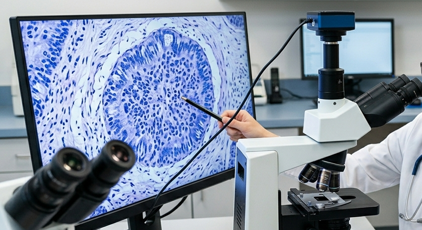
If basal cell carcinoma is suspected, the provider will usually perform a biopsy. This involves removing a small sample of the lesion, or sometimes the entire lesion, and sending it to a laboratory. A pathologist examines the tissue under a microscope to confirm whether cancer cells are present and identify the subtype.
Common biopsy methods include:
- Shave biopsy: A thin layer of tissue is removed from the surface.
- Punch biopsy: A circular tool removes a deeper sample.
- Excisional biopsy: The entire suspicious area is removed.
A biopsy is important because different skin cancers can look similar. Treatment planning depends on the diagnosis, size, location, depth, and microscopic features of the tumor.
Treatment Options
Basal cell carcinoma is highly treatable, especially when found early. The best treatment depends on the tumor’s size, location, subtype, depth, recurrence risk, and the patient’s overall health.
Surgical Excision
In surgical excision, the doctor cuts out the cancer along with a margin of normal-looking skin. The tissue is usually sent to a lab to check whether the margins are clear. This method is commonly used for many BCCs and has a high cure rate.
Mohs Micrographic Surgery
Mohs surgery is a specialized technique often used for BCCs on the face, ears, scalp, hands, genitals, or other areas where preserving healthy tissue is important. During Mohs surgery, the surgeon removes the cancer layer by layer and examines each layer under a microscope during the procedure. This continues until no cancer cells remain.
Mohs surgery is especially useful for recurrent tumors, aggressive subtypes, tumors with poorly defined borders, and lesions in cosmetically sensitive areas.
Curettage and Electrodesiccation
This method involves scraping away the tumor with a curette and using electric current to destroy remaining cancer cells. It may be used for certain small, low-risk BCCs, often on the trunk or limbs. It is generally not preferred for high-risk areas or aggressive subtypes.
Cryotherapy
Cryotherapy uses extreme cold, usually liquid nitrogen, to destroy abnormal cells. It may be considered for selected superficial lesions, but it is not appropriate for all BCCs.
Topical Medications
Some superficial BCCs can be treated with prescription creams, such as imiquimod or 5-fluorouracil. These treatments are applied over several weeks and require careful follow-up. They are generally used for low-risk superficial BCCs rather than deeper or aggressive tumors.
Photodynamic Therapy
Photodynamic therapy involves applying a light-sensitive medication to the skin and then activating it with a special light. This can destroy cancerous and precancerous cells. It may be used for certain superficial BCCs, depending on location and risk factors.
Radiation Therapy
Radiation therapy may be used when surgery is not possible or not preferred, especially in older adults or people with medical conditions that make surgery difficult. It may also be considered for certain tumors in challenging locations.
Targeted Therapy and Immunotherapy
For rare advanced cases, such as BCC that has spread or cannot be treated with surgery or radiation, systemic medications may be used. These include targeted drugs that affect the hedgehog signaling pathway, such as vismodegib or sonidegib. In some cases, immunotherapy may be considered.
What Happens If BCC Is Left Untreated?
Basal cell carcinoma usually grows slowly, but it does not simply disappear. Without treatment, it can continue to enlarge and invade nearby structures. On the face, this can lead to significant tissue destruction. For example, a BCC near the nose, eyelid, or ear can damage cartilage or interfere with normal function.
Although metastasis is rare, local invasion can still be serious. Larger tumors often require more extensive treatment and may leave more noticeable scars. Early treatment is usually simpler, more effective, and cosmetically better.
Prevention: How to Reduce Your Risk
The most important prevention strategy is reducing UV exposure. This does not mean avoiding the outdoors completely, but it does mean protecting your skin consistently.
Helpful steps include:
- Use sunscreen daily. Choose a broad-spectrum sunscreen with SPF 30 or higher. Apply it generously and reapply every two hours when outdoors, or after swimming or sweating.
- Wear protective clothing. Long sleeves, wide-brimmed hats, and UV-blocking sunglasses provide strong protection.
- Seek shade. UV rays are usually strongest between late morning and mid-afternoon.
- Avoid tanning beds. Indoor tanning increases the risk of skin cancer and premature skin aging.
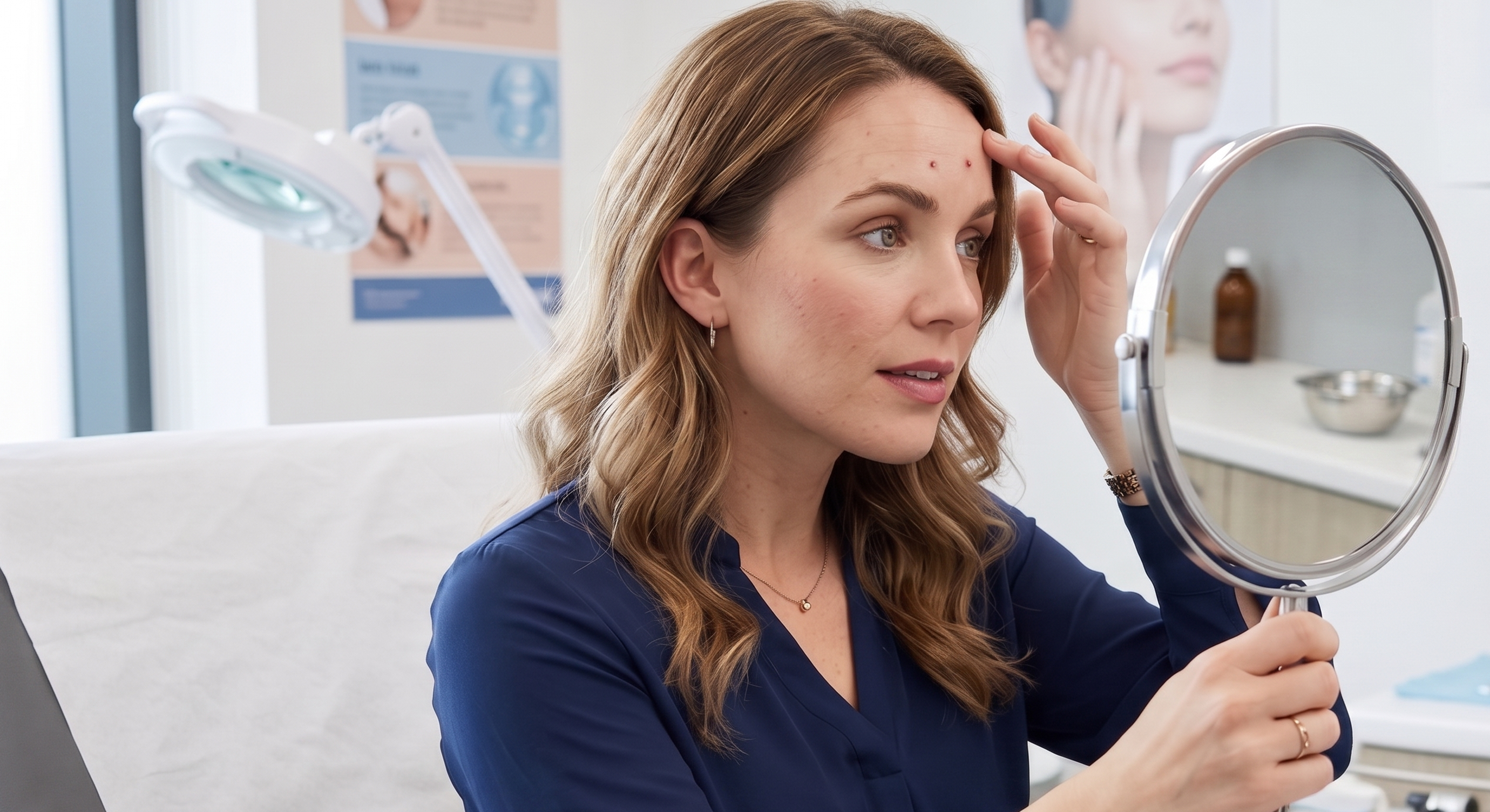
- Check your skin regularly. Look for new, changing, bleeding, or non-healing spots.
- Schedule professional skin exams. People with a history of skin cancer or high risk factors may need regular dermatologist visits.
Sun protection should start early in life, but it is never too late to benefit. Even after years of sun exposure, protecting your skin can reduce further damage and lower the risk of future skin cancers.
Living After a Basal Cell Carcinoma Diagnosis
A BCC diagnosis is usually manageable, but it can still cause anxiety. Many people worry about recurrence, scarring, or whether more cancers will appear. These concerns are valid. Having one basal cell carcinoma increases the chance of developing another, so follow-up care matters.
After treatment, your healthcare provider may recommend regular skin checks. The schedule depends on your personal risk, the type of BCC, and whether you have had multiple skin cancers. You should also watch the treated area for signs of recurrence, such as a new bump, persistent redness, bleeding, or a sore that does not heal.
It is also wise to become familiar with your own skin. Monthly self-exams can help you notice changes early. Use a mirror or ask someone you trust to check hard-to-see areas such as the back, scalp, and behind the ears.
When to See a Doctor
You should make an appointment with a dermatologist or healthcare provider if you notice:
- A sore that does not heal within a few weeks.
- A spot that bleeds, crusts, or repeatedly opens.
- A shiny, pearly, or translucent bump.
- A flat, scaly patch that persists.
- A scar-like area that appears without injury.
- A mole or dark spot that changes in size, shape, or color.
- Any skin lesion that looks unusual or concerns you.
Early evaluation can make treatment easier and more successful. Even if a spot turns out to be harmless, getting it checked can provide peace of mind.
Conclusion
Basal cell carcinoma is the most common skin cancer, but it is also one of the most treatable when detected early. It develops from basal cells in the skin and is most often linked to UV exposure from sunlight or tanning beds. While BCC rarely spreads to distant organs, it can grow into nearby tissue and cause damage if ignored.
Knowing the warning signs—such as a non-healing sore, shiny bump, scaly patch, or bleeding spot—can help you seek care promptly. Diagnosis usually requires a biopsy, and treatment options range from surgical removal and Mohs surgery to topical medications, radiation, and advanced therapies for rare cases.
The best defense is a combination of sun protection, regular skin checks, and timely medical care. Protecting your skin today can reduce your risk of future skin cancers and help keep your skin healthier for years to come.

Leave a comment
This site is protected by hCaptcha and the hCaptcha Privacy Policy and Terms of Service apply.